Structural and Mechanical Anisotropy in Human Cortical Bone Tissue
Graduate Student: Justin Deuerling

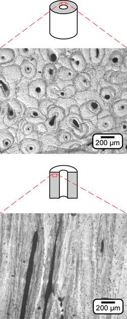
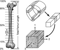
 Bone, as a biomaterial, consists of directional structural features across several unique hierarchical scales, ranging from nano-scale crystals and molecules to the macroscopic shape. However, the foundational structural unit across the hierarchical scales is a relatively simple two phase arrangement of anisometric bone mineral (apatite) preferentially oriented in a collagen matrix. Despite a growing database of measurements for the mechanical anisotropy of cortical bone, few efforts have been made to quantitatively measure influential structural features, e.g., the preferred orientation of bone mineral, and virtually no efforts have been made to correlate the anisotropy to structural measurements. Furthermore, the mechanical anisotropy in cortical bone is known to vary with anatomic location. Efforts to characterize and correlate structural features to mechanical anisotropy will also consider various anatomic sites, which will in turn provide data for clinically relevant anatomical sites (e.g., the proximal femur). The objectives of this work are to 1) characterize and quantitatively correlate anatomic variation in the mechanical anisotropy of human cortical bone with measurements of relevant structural features, 2) use this data to develop new micromechanical models which account for non-uniformity and anisotropy prior to hierarchical scaling, and 3) apply the knowledge gained in the design and synthesis of new orthopaedic biomaterials which can be tailored to function as a mechanical analog to human cortical bone.

