Scientists used to think that when a cell released a tiny, membrane-covered bubble—an extracellular vesicle (EV)—it was just taking out the trash. Then, in the 1990s and early 2000s, researchers discovered that these circulating microscopic bubbles were actually part of a critical communication network, carrying molecular instructions that affect how cells grow, divide, repair damaged tissue, respond to infection, and communicate with one another.
While these circulating EVs initially commanded the spotlight, another type of EV—one that stays put inside the tissue–has emerged as a potentially important player in the quest to engineer tissue to address age- and disease-associated dysfunction.
Pinar Zorlutuna, Roth-Gibson Professor of Bioengineering at the University of Notre Dame, and her lab have explored in an invited paper published in Nature Reviews Bioengineering that these matrix-bound EVs (MBVs) could be promising tools for regenerative medicine, tissue repair and disease diagnostics.
MBVs are tiny packages of biological information embedded throughout the body’s tissues. They carry proteins, RNAs and lipids that help tell neighboring cells how to behave. In cartilage, for example, MBVs can carry signals that suppress the growth of blood vessels—a critical function because healthy cartilage normally has no blood vessels, and abnormal vessel growth is associated with cartilage damage and disease.
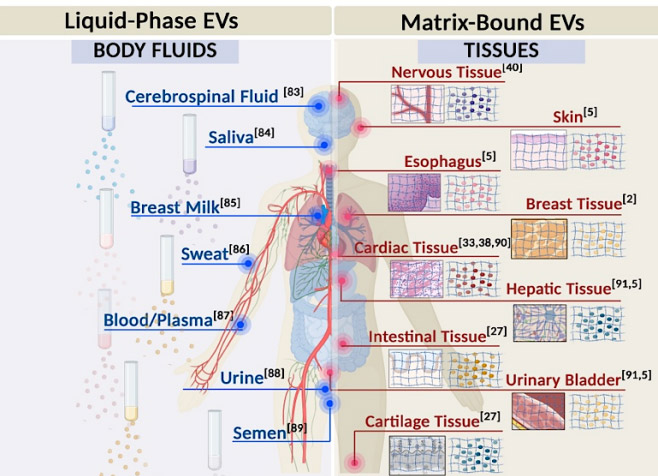
When breast tissue ages or hearts fail, MBVs molecular cargo can change in ways that promote inflammation, fibrosis or cancer-associated behavior. And this raises a compelling question: could health-promoting signals typically found in youthful MBVs be harnessed to treat disease? Researchers are exploring whether incorporating MBVs that carry potent tissue-protective and anti-inflammatory signals—or synthetic versions of them—into injectable hydrogels and medical scaffolds could improve cell health.
“Our lab is using what we know about MBVs to develop next-generation organ-on-a-chip models,” said Zorlutuna, referring to tiny laboratory versions of human tissues built with living cells. “Because MBVs carry tissue-, age-, and sex-specific signaling cues, incorporating them into these models could more accurately reflect the tissue’s microenvironment. When they’re combined with organ-on-chips and patient-derived cells, these models could provide a more personalized way to study disease and test potential therapies.”
Despite MBVs’ great promise, roadblocks remain. Since MBVs are interwoven with the extracellular matrix, developing easy ways to harvest them will be necessary before they can be incorporated into clinical practice. Also, healthy and diseased MBVs can look much the same on the outside. The differences may lie in the molecular messages they carry—and those messages can vary with age, sex, tissue location, and disease stage.
“Machine learning could help distinguish beneficial from pathological MBVs” said Zorlutuna. “AI can help scientists identify which molecules carried by these MBVs matter most, uncover patterns that signal health or disease, and that could accelerate the development of therapies.”
The Zorlutuna Lab operates at the intersection of tissue engineering, biomaterials, and cell biology. The lab focuses on designing biomimetic tissue models to understand complex cell-matrix interactions in heart disease, cancer, and aging, with the ultimate goal of engineering novel therapeutic platforms for tissue regeneration and repair.
—Karla Cruise, Notre Dame Engineering
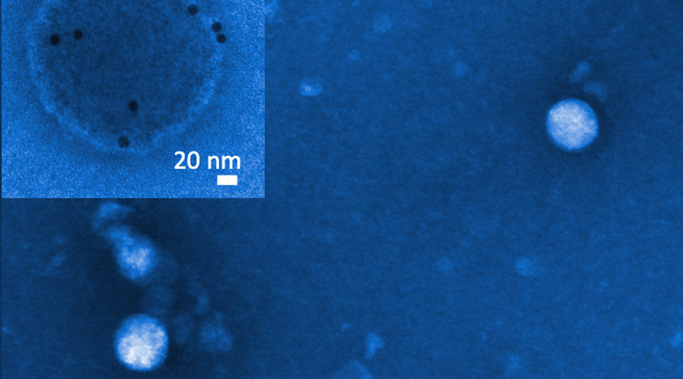
The image above, which appears in the article, is a microscopy view of matrix-bound vesicles (MBVs). While this process produces black-and-white images only, blue was added here to highlight MBV structures.